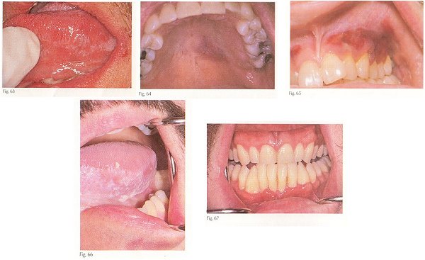
Candidiasis Colonization and infection of the oral mucosa by Candida albicans is among the earliest and most common findings in HIV-infected patients. In one study, 88% had oral candidiasis. Lesions range from white to red or red/ white combinations. Figure 63 illustrates the typical appearance of candidiasis on the lateral tongue; note the resemblance to hairy leukoplakia. The lesions may be asymptomatic or there may be mild discomfort.
Kaposi's sarcoma -AIDS patients are vulnerable to a variety of oral malignancies including Kaposi's sarcoma, malignant lymphoma and squamous carcinoma. Kaposi's sarcoma is the most common. In one study, 20% of AIDS patients had Kaposi's sarcoma and of these, the tumor was in the oral cavity in 1 of every 5 patients; the palate is the most common site. In the early stage, the tumor appears as a red to purple bruise (fig. 64). The tumor grows and eventually appears as a hemorrhagic mass (fig. 65). The cell of origin is endothelium; thus Kaposi's sarcoma is a variety of angiosarcoma. They are locally invasive, cause pain and bleeding and interfere with normal function. Radiation is the preferred treatment but laser resection and intralesional vinblastine provide palliation.
Hairy leukoplakia -This variety of leukoplakia was first recognized in HIV-infected patients but has been encountered in other immune deficiency states such as organ transplant patients who are intentionally immune suppressed. The lateral tongue is the most common location (fig. 66). lesions are of rough texture, adherent and asymptomatic. The diagnosis of hairy leukoplakia can be suspected on routine biopsy specimens, but confirmation requires demonstration of the presence of the causative virus, the Epstein-Barr herpesvirus. This is ordinarily achieved by DNA in situ hybridization. A word of caution: hairy leukoplakia may be confused with candidiasis. In one study, 52% of cases clinically diagnosed as hairy leukoplakia proved to be candidiasis. A patient who presents with a white lesion should be treated with antifungal therapy first. If it fails to heal, it most likely is hairy leukoplakia.
Gingival and periodontal lesions -HIV infected patients are vulnerable to gingivitis, periodontitis, (fig. 67) and necrotizing ulcerative gingivitis (ANUG-like). The organisms recovered from these lesions are the same as those in non-HIV-positive patients, but they are present in greater numbers. Lesions are treated by dental phophylaxis, debridement, and metronidazole. Good oral hygiene and daily rinses with chlorhexidine are beneficial.
Others -HIV patients also develop major aphthous-like lesions that respond to topical tetracycline and topical steroid therapy. Other patients have painful palatal and gingival ulcers that have been found to harbor cytomegalovirus. The human papillomavirus has also been found in mucosal papules. Herpesvirus may cause painful and protracted oral ulcers that are responsive to treatment with acyclovir. Lastly, xerostomia secondary to a Sjogren's syndrome-like illness has been reported. |