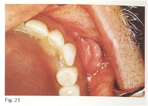
Description: This lesion occurs in those who wear dentures. The lesion consists of two or more folds of soft tissue separated by a central groove into which fits the denture border (Fig. 23). It most often is found in the buccal vestibule of the anterior maxilla but any mucosal area touched by a denture border is vulnerable including the lingual aspect of the mandible. In a study of 583 cases, 64% were found in females. Those in the fifth and sixth decade are most often affected. Duration ranged from 1 week to 10 years, 40% of the patients reported a duration of 6 months to 2 years. Symptoms are absent except in ulcerated lesions, which may be painful. Histologically the excessive tissue is composed of cellular, inflamed fibrous connective tissue.
Etiology: This is an inflammatory fibrous hyperplasia of oral mucosa caused by an over-extended denture border.
Treatment: Surgical excision of the lesion and reduction of the denture border.
Prognosis: Good
Differential diagnosis: The lesion has such a characteristic clinical appearance that differential diagnosis is not a problem. In questionable cases, biopsy should be done. . |